 |
Last Update:
Thursday November 22, 2018
|
| [Home] |
|
Volume 18 Issue 1 Pages 1 - 53 (April 2001) Citation: Jansman, J., Chanin, P.R.F. & Dallas, J.F. (2001) Monitoring otter populations by DNA typing of spraints IUCN Otter Spec. Group Bull. 18(1): 12 - 19 Monitoring otter populations by DNA typing of spraints Hugh A.H. Jansman1, Paul R.F.Chanin2 and John F. Dallas3 1 Alterra.
Department of Ecology and Environment, P.O. Box 47, 6700 AA Wageningen,
The Netherlands
E-mail: h.a.h.jansman@alterra.wag-ur.nl (received 6th April 2001, accepted 21st May 2001)
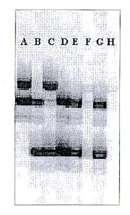
INTRODUCTION Habitat loss has created fragmented populations in many species of animals. Such populations can suffer from reduced mating opportunities and inbreeding depression. Conservationists have responsibilities to monitor such species to ensure that populations remain viable. Unfortunately, such species are usually elusive and their numbers low, so monitoring by direct observation is often difficult or impossible. The need for an alternative monitoring method has prompted scientists to develop genetic typing of DNA extracted from animal droppings (molecular scatology). Droppings of many species can be collected easily and in some cases they provide the only evidence that a species exists. Conservationists who attended the VIII International Otter Colloquium in Valdivia, Chile, 2001, expressed great interest in molecular scatology. The aim of this article is therefore to give guidelines based on two feasibility studies on how to apply this method to the Eurasian otter Lutra lutra and other otter species. Two groups in Europe have experience of DNA typing of L. lutra spraints. A feasibility study in the UK was done to assess this method for surveying wild L. lutra. The study was a joint effort involving the Environment Agency, the Universities of Aberdeen and Exeter, the Somerset Otter Group and the Devon and Hampshire Wildlife Trusts, together with many volunteers, without whom the study would have been impossible. Over 600 spraints were collected from four river catchments during 18 months and analysed. The study is published (COXON et al, 1999) and summarised at www.ex.ac.uk/mammals/dna/. The Alterra group subsequently acquired the DNA typing method from the Aberdeen group to monitor the success of the future reintroduction of L. lutra in The Netherlands. Hair and spraint samples were collected from captive otters at the breeding station Aqualutra and Ouwehands Zoo, and reference samples of Scottish otters were included. DNA TYPING Spraints are easy to collect because otters deposit them at highly visible points near waterway margins. Spraints contain the DNA of their previous owner in the form of cells shed from the intestinal lining. In DNA typing, the owner is identified according to a genetic profile consisting of 6-15 highly variable microsatellites and one male-specific gene. The profiles are detected using the polymerase chain reaction (PCR). PCR primers for 13 L. lutra microsatellites and a male-specific L. lutra gene are available. These primers detect only otter DNA, not DNA from gut bacteria or prey species. Some of the microsatellite primers and the male-specific primer are equally applicable to representative species of all four otter genera (DALLAS and PIERTNEY, 1998; DALLAS et al., 2000). DNA typing of wild L. lutra populations and other otter species could meet with two obstacles. First, populations with a long history of low numbers, reintroduced populations stemming from few founders and geographically isolated populations, could contain so little microsatellite polymorphism that many individuals will have identical genotypes. Good examples are the L. lutra population on the island of Shetland, UK, which contains less than half the level of microsatellite polymorphism found in populations in mainland Scotland (DALLAS et al., 1999), and the reintroduced L. lutra population in the River Itchen catchment in southern Britain (COXON et al., 1999). Second, some of the available L. lutra microsatellites could have low polymorphism in other species. Developing more microsatellites for L. lutra and other otter species could overcome both obstacles. Spraint DNA typing has three parts. First, DNA is extracted from spraints. Because the quality and quantity of such DNA are typically low, DNA recovery must be efficient and PCR inhibitors must be excluded. Second, the otter microsatellites and SRY gene are amplified to yield detectable PCR product. Third, PCR products of different length are separated by electrophoresis and detected (Fig. 1). The resulting genetic profiles are subjected to population genetic analysis using computer programs. Extraction of DNA from spraints has been done by two methods. The Aberdeen group used a CTAB/GITC/DIATOM/ VECTASPIN method (PARSONS et al., 1999) whereas Alterra used a commercial kit designed for DNA extraction from animal tissue (QIAgen; www.qiagen.com). A direct comparison of the PCR success rates of these methods remains to be done. Detection of PCR products was done using two systems. The Aberdeen group used standard sequencing gels and autoradiography whereas Alterra used a Licor DNA sequencer/fluorescence system. The Licor/fluorescence system is generally more sensitive than autoradiography and thus superior for spraint DNA analysis. The Aberdeen group obtained a success rate of around 20% for typing at least seven out of nine microsatellites and the SRY gene, whereas Alterra obtained an approximately three-fold higher rate (50-60%; H. Jansman, unpublished) for typing 5 microsatellites and the SRY gene. Although the differences in DNA extraction and detection methods probably contributed to the difference, another important factor was probably the age and type of spraints. At Alterra, a few very fresh (approximately 1-6 hours old and immediately extracted) spraints from zoos were analysed whereas in the UK study over 600 wild-collected spraints that averaged 12 hours old were analysed. The success rate of DNA typing wild-collected spraints at Alterra could therefore be lower than 50%. We expect to find out if this is the case during summer 2001, when spraints from the Trebon population in the Czech republic will be analysed at Alterra. A commercial company in the UK specialising in DNA analysis (Tepnel; www.tepnel.com) recently carried out a small study to improve the success rate of spraint DNA typing. It was found that a 504 bp portion of the mitochondrial DNA gene cytochrome B was detectable in approximately 80% of spraint DNA extracts. The success rate of microsatellite typing of these samples by multiplex PCR is presently being assessed. Commercial laboratories could therefore provide services for acquiring raw genetic data. University or research institutes could also provide the skills necessary for genetic data analysis and ecological interpretation. More detailed protocols and links will appear on the website: http://www.ex.ac.uk/mammals/dna/. INFORMATION FROM SPRAINT DNA TYPING Low-intensity sampling and spraint DNA typing could yield data to distinguish most unrelated individuals present and estimate levels of genetic diversity in the population. High-intensity spraint sampling of populations containing high levels of microsatellite polymorphism could also yield estimates of individual home ranges and dispersal patterns. In areas where more than one otter species occurs, spraint DNA typing could also identify species. This method can distinguish among L. lutra, American mink Mustela vison and polecat Mustela putorius (HANSEN and JACOBSEN, 1999), and DNA sequences suitable for designing species-specific PCR primers for otters are available (KOEPFLI and WAYNE, 1998). Spraints also contain food remains and parasites, perhaps allowing diet and disease analysis by PCR (KOHN and WAYNE, 1997). Lastly, the reproductive status of individual otters (separation of juveniles/sexually-inactive females, pregnant females and adult males) can be studied by analysing hormone metabolites in otter spraints (TSCHIRCH et al., 1996). PRACTICAL TIPS We emphasise that otter DNA extracted from spraints is generally of poor quality and yields low typing success rates and high rates of potential errors in individual profiles (TABERLET et al., 1999). It is therefore necessary to standardise the practical aspects of the method. Two important factors under user control are the age and storage conditions of spraints. Spraints should be collected as soon as possible after deposition. In the UK study, spraints were collected during the early morning and only those judged by then-fresh appearance to have been deposited during the previous night were collected. The success rate appeared higher for anal jelly samples than for normal spraints (COXON et al., 1999), and other analyses of spraints from captive otters suggested that typing success rate decreases from 60% at deposition to 15% after 24 hours (J. Dallas, unpublished). In confirmation, the Alterra group found very low success rates in typing of spraints over three days old (H. Jansman, unpublished). For monitoring studies, we strongly recommend collecting only fresh (<12 hours old) spraints, preferably before 10 a.m. in the morning. When spraints are rare and otter numbers are suspected to be low, it could be worth collecting older spraints, despite a much lower success rate. Spraint storage conditions are important because correct storage retards or stops DNA breakdown. Ideally, a spraint (or a part of it) should be stored in a plastic vial containing a suitable solution, and DNA should be extracted as soon as possible. Suitable storage solutions are 95% ethanol and ATL buffer from the animal tissue DNA extraction kit (QIAgen). Irrespective of which storage solution is used, any storage of samples should be done at -20°C. Under less optimal field conditions, it could be effective to preserve DNA in spraints by drying with silica beads (WASSER et al., 1997). All samples should be well-documented (date, location, composition) using pencil to write on the vials. Before starting a monitoring program based on spraint DNA typing it is important to verify that levels of microsatellite polymorphism in the population are high enough, at least for individual identification. A representative sample of 10-20 otters from the population should therefore be analysed by DNA typing. Suitable samples are hair (>30 for each individual, including roots), blood, or tissue from dead otters. The UK study showed that when average expected heterozygosity per locus (Hc) is below 0.40, and the average number of alleles per locus (Nall) is below 3.0, even unrelated individuals can have identical genotypes for up to nine microsatellites. In this case, population size can be underestimated owing to this "shadow effect" (MILLS et al., 2000) and home range sizes overestimated. The number of microsatellites used, and levels of polymorphism of each locus, is also important. The more loci are typed, the more likely different individuals can be discriminated, but the more likely false genotypes will arise through typing error. Six microsatellites (Lut 701, 715, 717, 832, 833 and 902; DALLAS and PIERTNEY, 1998; DALLAS et al., 1999) were used for spraint DNA typing in the UK study, and a further three (Lut 435, 457, 615) were typed subsequently (J. Dallas, unpublished). In highly polymorphic populations (H > 0.6, Nall > 4.0) the use of six loci is probably sufficient for individual identification and monitoring levels of genetic diversity. Intensive individual-level analyses, and any analyses of genetically depauperate populations will require 10-15 loci. It is essential that all DNA samples be typed up to seven times (TABERLET et at., 1996; 1999; BAYES et al., 2000; GOOSSENS et al., 2000). Only individual profiles that are consistent in such replicate typing should be considered reliable. When the profiles of two spraints match, it is important to calculate ratio of the probabilities that they represent the same individual or two individuals having a given level of relatedness. Lastly, standardisation of methods among groups will be important for reliable comparisons of DNA typing data. CONCLUSION AND RECOMMENDATIONS Spraint DNA typing shows promise as a tool for monitoring of otter populations. This method can already identify different individuals and thereby provide estimates of sex ratio and minimum population size. Further progress will depend on achieving higher success rates, lower cost, and developing more highly variable microsatellites and species-specific PCR assays. One recommendation from the IUCN otter specialist group to participants at the VIII international otter colloquium in Valdivia, Chile was to document the distribution and status of all 13 species of otters globally. The use of DNA typing in endangered and poorly known otter species could make an important contribution to this goal. We therefore recommend that skin, tissue or DNA samples from all endangered otter species be archived for future genetic analysis. ACKNOWLEDGEMENTS - We thank Aqualutra and Ouwehands Zoo for access to their captive otters and all the volunteers, without whom the study would have been impossible REFERENCES Bayes, M.K., Smith, K.L., Alberts, S.C.,
Altmann, J. & Bruford, M.W. 2000. Testing the reliability of
microsatellite typing from faecal DNA in the savannah baboon. Cons.
Genet. 1, 173-176. Résumé : Étude des Populations de
Loutres Grâce au Test ADN sur les Épreintes Resumen: Monitoreo de Nutrias Mediante
Identificación de Fecas por ADN |
| [Copyright © 2006 - 2050 IUCN/SSC OSG] | [Home] | [Contact Us] |